
Advanced medical imaging technology has provided remarkable new insights into the lives of ancient Egyptians, revealing that individuals from over two millennia ago suffered from health conditions strikingly similar to those affecting modern populations.
Groundbreaking Analysis of Preserved Remains
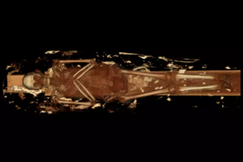
Radiologists from the Keck Medicine team at the University of Southern California have conducted detailed computed tomography scans on two linen-wrapped mummies dating from 330BC and 190BC. The non-invasive examination technique has allowed researchers to peer beneath the surface of these ancient remains without causing any damage to the precious specimens.
Revealing Ancient Health Conditions
The comprehensive analysis uncovered specific health challenges faced by these individuals during their lifetimes. The elder of the two mummies displayed clear evidence of spinal degeneration, with a collapsed lumbar vertebra in the lower back region. This condition, likely resulting from natural aging processes and physical wear and tear, would have caused significant discomfort and mobility issues.

The second individual presented different health concerns, including substantial dental problems and severe deterioration of the hip joint. Researchers determined this person was older at the time of death, with the scans providing detailed information about their physical condition that would have been impossible to obtain through traditional archaeological methods.
Technological Advancements in Archaeological Research
Dr Summer Decker, who leads 3D imaging initiatives for Keck Medicine, explained the significance of the technological progress: "These mummies were scanned previously, but due to advancements in scanning technology, the results are much more detailed and extensive than ever before." The new high-resolution images have revealed previously unknown details that help construct a more complete picture of ancient Egyptian life.
The CT scanning process creates hundreds of detailed cross-sectional images that experts can digitally assemble into comprehensive three-dimensional models. This technology, commonly used in modern surgical procedures, has revolutionised the study of ancient specimens by allowing thorough examination without physical disturbance.
Cultural Context and Personal Details
Beyond medical information, the scans have provided insights into cultural practices and personal characteristics. The mummy with spinal issues was buried with several artefacts representing scarab beetles and a fish, offering clues about burial traditions and symbolic meanings in ancient Egyptian culture.
Researchers were also able to reconstruct facial features with remarkable precision, including the shapes of eyelids and lower lips. This level of detail brings these ancient individuals to life in ways previously unimaginable, connecting modern observers directly with people from a distant past.
Educational Exhibition and Future Research
The mummies, along with their corresponding 3D digital models and research findings, will be featured in an upcoming exhibition at the California Science Center. Diane Perlov, senior vice president for special projects at the institution, expressed enthusiasm about the discoveries: "Seeing beneath the surface to reveal the specific lived experience of individuals is incredibly exciting. This modern scientific technology offers us a powerful window into the world of ancient people and past civilisations that might otherwise be lost."
The research demonstrates how medical imaging technology originally developed for clinical applications has found valuable applications in archaeological and historical research. As scanning technology continues to advance, researchers anticipate even more detailed revelations about ancient civilisations and their inhabitants.
This intersection of medical science and archaeology provides not only historical insights but also valuable perspectives on human health across millennia, showing how certain physical conditions have remained constant challenges throughout human history.