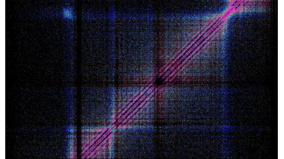
British researchers from the Universities of Oxford and Cambridge have produced the most detailed image of the human genome ever captured, offering unprecedented insights into how DNA functions inside cells. The breakthrough, published in the journal Cell, could pave the way for new treatments for diseases such as cancer and Alzheimer's.
For over two decades, scientists have known the complete sequence of the human genome—three billion 'letters' of DNA—but how this genetic code folds and operates as a three-dimensional structure remained elusive. Each cell contains about two metres of DNA compressed into a space just one-hundredth of a millimetre across, constantly bending and looping to bring distant sections into contact.
This intricate structure determines which genes are active or silent, akin to a circuit board controlling switches. Lead author Professor James Davies of Oxford University said: 'For the first time, we can see how the genome's control switches are physically arranged inside cells. This changes our understanding of how genes work and how things go wrong in disease.'
The new technique, called MCC ultra, allowed scientists to visualise DNA down to a single-base pair—the smallest unit of DNA—200 times better than previous methods. This revealed how cells use electromagnetic forces to bring DNA control sequences to the surface, clustering into 'islands' of gene activity. Researcher Hangpeng Li noted: 'We now have a tool that lets us study how genes are controlled in exquisite detail. That's a critical step toward understanding what goes wrong in disease, and what might be done to fix it.'
Most common genetic changes linked to diseases do not alter genes themselves but affect which aspects are switched on or off. The research, funded by the UK's Medical Research Council, represents a major advance in molecular genetics, shedding light on conditions like heart disease, autoimmune disorders, and cancer.