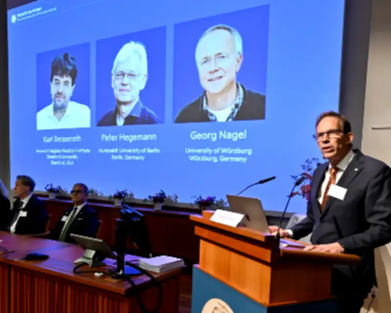
The 2026 Nobel Prize in Physiology or Medicine has been awarded to three scientists for their research into the brain's workings. Karl Deisseroth of the Howard Hughes Medical Institute and Stanford University, Peter Hegemann of the Humboldt University of Berlin, and Georg Nagel of the University of Würzburg received the prize for their discoveries concerning light-gated ion channels and optogenetics.
The Nobel assembly at the Karolinska Institute in Stockholm, Sweden, announced the winners on Monday. They will each receive an equal share of a prize of 12m Swedish kronor (about £900,000).
117th Award and Historical Context
The 2026 award marks the 117th time the prize has been given. Of the 235 laureates – including the latest winners – only 14 have been women, and none have been awarded to black scientists.
The trio's research led to the development of a light-sensitive switch that can be used to control nerve cells in the living brain, switching them on or off.
Origins in Algae and Channelrhodopsins
The discovery originated in research by Hegemann, who explored how a green alga called Chlamydomonas swam towards light. Abdel El Manira, a professor of neuroscience at the Karolinska Institute and member of the Nobel assembly, explained that Hegemann discovered the alga's eye spot contains a light-sensitive protein that converts light almost instantly into an electrical signal, allowing the alga to move.
Nagel and Hegemann identified the proteins, called channelrhodopsins. Working with frog egg cells, they revealed that when the proteins were exposed to blue light, they allowed charged particles – or ions – to flow into the cell, producing an electrical impulse. Further work revealed that when the genetic instructions for one of the proteins were inserted into human or mouse kidney cells, those cells became light sensitive.
From Rats to Mice and Medical Applications
Deisseroth took the discovery further, using rats to demonstrate that introducing the gene for a channelrhodopsin into nerve cells made it possible to produce electrical signals in them using light. He and his colleagues later applied the approach to nerve cells in the motor cortex of mice brains, using light delivered by tiny optic fibres passed through their skull to trigger movement in the rodents' whiskers. Subsequent work in mice showed the approach could also investigate which networks of nerve cells were involved in forming particular memories, including fear.
The work has opened up a field of medicine known as optogenetic therapy. In one trial, it was used to partially restore the sight of a man who went blind from retinitis pigmentosa, a condition that damages light-sensitive cells in the retina. The genetic instructions for making a channelrhodopsin were delivered by a harmless virus into retinal ganglion cells in the man's eyes. He was then fitted with light-stimulating goggles that captured pictures of the world around him and converted them into single-wavelength images projected on to his retina using pulses of light.
Reactions and Future Potential
Prof Botond Roska, the director of the Institute of Molecular and Clinical Ophthalmology Basel who co-led the study, welcomed the announcement. “This is truly wonderful news. I hope this helps to boost all efforts on optogenetic vision restoration to bring back vision to blind patients,” he said.
Anna Wedell, a professor at Karolinska Institute and a member of the Nobel committee for physiology or medicine, said the laureates' work provided new possibilities to map which cells were responsible for which functions and which cells communicated with each other, opening up a new understanding of brain function. “The brain is our most complex organ and we have very little understanding of how it works and we have had very few methods to proceed in that understanding,” she said. “We've had anatomical maps and correlations, but now we have a tool that can provide cause-and-effect relationships so we can get a functional map of the brain, exactly pinpoint which cells do what, which cells are responsible for creating behaviours or even emotions and memories in the brain.”
Wedell said that by studying animals to understand how a normal, healthy brain worked, it was then possible to explore what happens when things go wrong – for example in diseases such as dementia, epilepsy and addiction. “By understanding which cells are active in these diseases in the mouse, we can also understand where to look in the human,” she said.