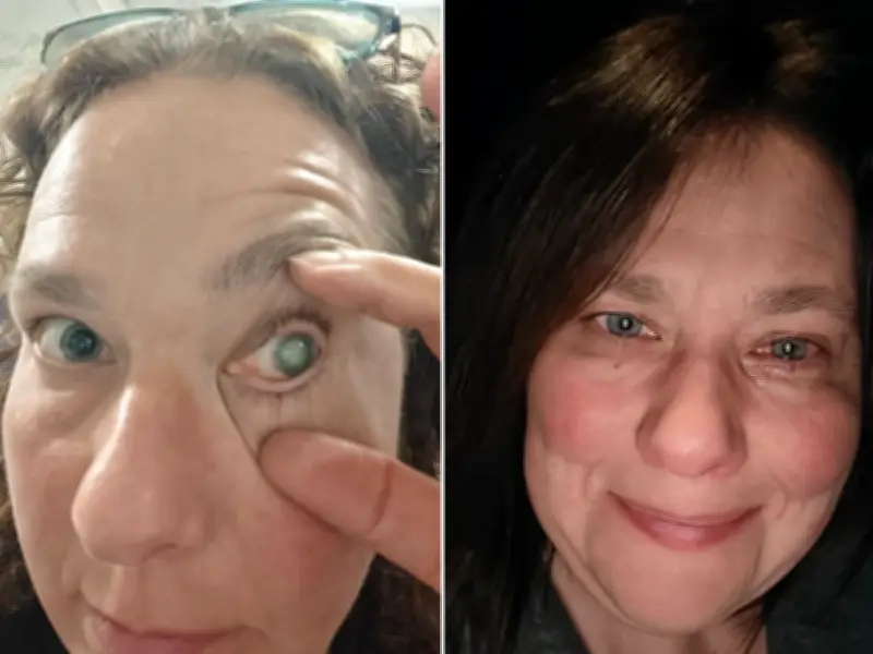
Claire Buxton, 46, from the Midlands, is set to have her left eye removed after a rare parasitic infection linked to contact lens wear destroyed her vision. She woke up with what felt like a grain of sand in her eye, dismissed it as conjunctivitis, but within weeks her sight deteriorated dramatically. She was diagnosed with Acanthamoeba keratitis (AK), a rare infection caused by microscopic parasites that can become trapped under contact lenses.
Infection and misdiagnosis
Claire wore daily and two-week contact lenses from morning until night, occasionally showering in them. She believes she contracted the infection after playing field hockey on a water-based artificial grass pitch. The day after the game, she experienced redness and discomfort. Pharmacies treated her for conjunctivitis, but her condition worsened. Referred to an eye unit, she was initially misdiagnosed and prescribed steroid eye drops, which experts say can suppress the immune response and worsen the infection.
After a corneal scrape, Claire was diagnosed with AK and began intensive treatment: three different anti-parasitic eye drops every hour, alongside antibiotics, lubricants, and oral steroids. Her vision now fluctuates between reading a few lines on an eye chart and perceiving only light.
Decision for removal
Specialists were divided between a risky corneal transplant and eye removal with a prosthetic. After a year, Claire chose removal. “I was still offered a transplant as an option, but the risks involved seemed to outweigh the benefits. I can have my eye removed and a hand-painted prosthesis to match my other eye. It'll mean no more pain, no more worrying about dust or water getting in my eye,” she said.
According to Moorfields Eye Hospital, about two in 100,000 contact lens wearers per year in the UK are diagnosed with AK. The infection is caused by a microscopic organism found in bodies of water, tap water, swimming pools, hot tubs, soil, and air. While some recover in three to six months, complicated cases often lead to surgery and permanent sight loss.
Second case highlights delays
Charlie Shields, 26, from Chiswick, west London, suffered weeks of escalating pain and repeated misdiagnoses. His eye became red after work, then developed extreme light sensitivity and pain. Doctors initially diagnosed inflammation and later ocular herpes, prescribing steroid drops that allowed the parasite to spread deeper into his cornea. A corneal specialist finally confirmed AK with specialised imaging.
“They told me not to Google it. Then they said I had to stop everything I was taking and start putting disinfectant eye drops in every hour, day and night. The steroids I'd been using had actually fed the parasites and made them burrow deeper into my eye,” Charlie recalled. For two weeks, he woke every hour to administer painful drops, leaving him virtually sleepless. He has now had more than 5,000 eye drops.
Charlie rarely wore contact lenses and had no obvious risk factors. “I'd never even heard of this disease. I wasn't wearing lenses in the shower or swimming, and I only wore them occasionally. I was as careful as the next person, but now I realise people don't understand how serious this infection can be,” he said. His vision remains permanently affected, resembling “looking through frosted glass”.
Importance of early diagnosis
Early symptoms of AK include persistent redness, disproportionate pain, blurred vision, excessive watering, and extreme light sensitivity. Specialists stress that prompt diagnosis is crucial, as delays can lead to permanent sight loss or the need for corneal transplantation. Claire continues to wear a contact lens in her good eye for hockey, but is now much more careful. “I'm processing the loss of hope, grieving the trauma and the restrictive life and pain I've had to endure over the last 18 months. I'm glad to be able to put it behind me and move on,” she said.